Cornea of Eye
What is the Cornea?
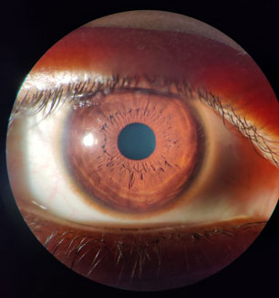
The cornea is the outermost transparent layer of the eye that covers the pupil and the iris. It is comparable to the clear glass placed over a watch dial, protective, yet crystal clear.
So what is cornea, exactly? It's the eye's clear "window." Its primary function is to help the eye focus, by bending light rays onto the retina at the back of the eye, the cornea allows us to see clearly and sharply.
Dr Kareeshma Wadia is one of the most trusted cornea specialist in Mumbai, she regularly sees patients with concerns ranging from blurry vision to corneal infections, many of which trace back to changes in this thin, delicate layer.
Anatomy of the Cornea
- Layers of the Cornea
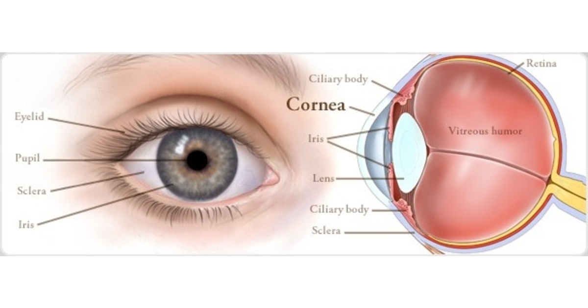
Understanding cornea anatomy helps explain why even small injuries or infections can affect vision. The cornea is made up of five main layers:
Epithelium: The outermost layer, acting as a barrier that keeps foreign objects out of the eye.
Bowman’s layer: A thin layer joining the epithelium and the stroma.
Stroma: The thickest layer of the cornea, responsible for giving it its dome shape.
Descemet’s membrane: A thin layer separating the stroma from the endothelium.
Endothelium: A single layer of cells sitting between the stroma and the aqueous humor (the clear, jelly-like fluid inside the eye).
A sixth layer, known as Dua's layer, was more recently discovered and has since been added to our anatomical understanding of the cornea.
- Corneal Shape and Transparency
Much like the glass on a watch dial, the cornea is clear and dome-shaped, a structure that is essential to its role in vision.
Function of the Cornea
The cornea works like a camera lens, focusing incoming light onto the retina so we experience clear, sharp vision. In fact, the cornea accounts for the majority of the eye's total focusing power.
Acting as a shield, the cornea prevents dust, debris, and other foreign objects from entering or damaging the inner eye.
How Are Cornea Problems Diagnosed?
A cornea specialist or eye doctor will typically carry out a thorough eye examination, which may include:
Common Corneal Conditions
1. Corneal Infections (Keratitis)
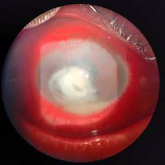
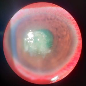
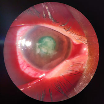
Corneal infections, commonly known as keratitis, occur when bacteria, viruses, or parasites affect the cornea. The most frequent cause is overuse of contact lenses, including sleeping with lenses in, or using homemade, non-sterile lens solutions.
Symptoms of corneal infections can include:
Treatment: Anyone experiencing these symptoms should see an ophthalmologist immediately to prevent further damage or scarring. Once diagnosed, corneal infections are treated with antibiotics or other appropriate medication depending on the cause.
The term "keratitis" refers broadly to infections or immune reactions affecting the cornea, while a "corneal ulcer" describes an infection that has progressed deep enough to break down the cornea's intact layers. Both are usually accompanied by a loss of corneal transparency, which is why blurred vision is often the first complaint. Healing tends to take longer once infection sets in, and the right anti-infective drops or oral medication are used to stop it from spreading. In some cases, keratitis heals with scarring that leaves behind persistent blurry vision, and in severe cases, a cornea transplant may become necessary.
2. Corneal Dystrophy and Degeneration
Corneal dystrophy conditions are relatively rare, can affect one or both eyes, and are often idiopathic (with no identifiable cause). They tend to recur even after surgical treatment. More than 20 types of corneal dystrophies exist, most of them hereditary and slow-progressing. The most common include:
1. Fuchs' Dystrophy: The cornea swells, becomes painful, and vision becomes distorted. Management: If saline drops, soft contact lenses, or ointments don't control the swelling, and the condition advances quickly, a cornea transplant may be required.
2. Granular Dystrophy: Causes lesions resembling granules or crumbs inside the eye, which can become painful and affect vision over time.
3. Lattice Dystrophy: Abnormal protein fibres build up in the stroma, gradually clouding the cornea and affecting vision. Management: In severe cases, a corneal transplant restores eyesight. Many patients develop corneal scarring that causes persistent haze, which can worsen over time. A laser-based procedure called photothermal keratectomy (PTK) may also be considered if scarring develops on the donor tissue post-transplant.
4. Map-Dot Fingerprint Dystrophy: The epithelium doesn't form normally and erodes, altering the cornea's curvature and sometimes causing blurred vision. Management: Early stages are treated with eye drops or ointments. Corneal scraping and PTK are additional options for relief.
3. Corneal Injuries
Corneal injuries are broadly classified as traumatic or exposure-related.
Traumatic injuries: include scratches, abrasions, or a foreign body entering the eye, such as a scratch from a fingernail or an object like a pencil accidentally striking the eye.
Exposure-related injuries: result from burns caused by heat, chemicals, or radiation.
Corneal Transplantation
A cornea transplant may be necessary when the cornea has suffered significant damage from injury, or when an existing corneal dystrophy has progressed and worsened.
During the procedure, the damaged layer of the cornea is removed in a circular pattern (similar to using a cookie cutter) and replaced with healthy donor tissue.
Full recovery from a cornea transplant can take up to a year. Most patients return to their regular routines within one to two weeks, with the exception of heavy lifting, which should be avoided for at least four weeks, or longer if advised by your doctor.
Dr Kareeshma Wadia completed a super-specialisation in this field, spending two years at Narayana Nethralaya Superspeciality Eye Hospital in Bangalore to master cornea transplant surgery, making her one of the more experienced choices for patients seeking a cornea specialist in Mumbai.
Specular Microscopy
Specular microscopy is a non-invasive photographic technique used to visualize and analyze the corneal endothelium, the innermost layer of the cornea responsible for keeping it healthy and clear.
This scan checks the number of endothelial cells present in the cornea. A healthy cornea typically has hexagon-shaped endothelial cells at a density of around 3,000 cells; in a compromised or diseased cornea, cell shape becomes irregular and density can fall below 1,000. This test helps an eye specialist diagnose Fuchs' Dystrophy and other conditions affecting the endothelium.
Cornea Care and Hygiene
1. Schedule regular checkups with your eye doctor, which may include specular microscopy
2. Wear sunglasses to reduce UV exposure
3. Monitor your eye health alongside conditions like diabetes
1. Always wash your hands before handling contact lenses
2. Follow the CRD method: Cleanse, Rinse, Disinfect (and dry) with the right solution
3. Never sleep with contact lenses on
4. Never clean lenses with saliva, tap water, or any non-sterile homemade solution
Lifestyle and Corneal Health
Eat a well-balanced diet rich in vitamins, minerals, zinc, copper, and vitamins A, E, and C.
Wear sunglasses to shield your eyes from UV radiation, and prioritise a good night's sleep.
Blinking regularly and moving your eyes up, down, and sideways are simple, effective exercises for eye health.
Visit our YouTube channel, "Dr Kareeshma Wadia — Jehan Eye Clinic," for a video guide to eye exercises.>
Why Corneal Health and Regular Eye Check-Ups Matter
The cornea is the very first layer of the eye, it acts as a barrier and protects the inner eye, playing a vital role in vision. Regular eye checkups, protective eyewear during sports, and sunglasses for UV protection all go a long way in preserving corneal health. Simple eye exams can even help detect underlying conditions like diabetes.
Why Choose Jehan Eye Clinic for Cornea Care
Jehan Eye Clinic is a trusted eye clinic in Vile Parle, Mumbai, combining modern ophthalmic technology with a patient-first approach. Led by Dr Kareeshma Wadia, an experienced cornea specialist in Vile Parle, the clinic offers evidence-based diagnosis and treatment for corneal infections, dystrophies, injuries, and transplants.
Whether you're searching for a reliable eye doctor in Mumbai, or a dedicated cornea specialist, Jehan Eye Clinic is committed to safe, ethical, and personalised eye care for patients of all ages.
Frequently Asked Questions

Jehan Eye Clinic is a center for skilled clinical excellence, and aims at being at par with the latest technology at all times. It is headed by Dr Kareeshma Wadia-Havewala, who is a specialist in Cataract, Cornea and Refractive Surgeries.
Quick Links
Contacts
Opening Hours
- Monday — 10am – 7pm
- Tuesday — 10am – 7pm
- Wednesday — 10am – 7pm
- Thursday — 10am – 7pm
- Friday —10am – 7pm