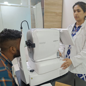
Optical Coherence Tomography (OCT Scan) is a non-invasive scan done as a diagnostic procedure in the clinic, which uses light rays to take cross sectional pictures of the different structures of the eye.
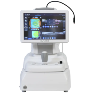
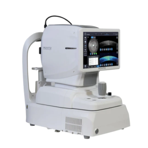
At Jehan Eye Clinic, we have the Huvitz OCT which can give us cross sectional scans as well as 3D images of the Retina, the Optic Nerve (which is affected in Glaucoma) and the Cornea.
RETINA
OCT of the retina can be done to diagnose and treat many disorders of the retina such as:
Macular Edema – this is the swelling of the retina which may happen in conditions such as Diabetes
Macular hole – the central part of the retina develops a hole that can start affecting the patient’s vision
Age related Macular Degeneration
Central Serous Chorioretinopathy
GLAUCOMA
OCT of the optic nerve is done to give an objective analysis of the Retinal Nerve Fibre Layer that surrounds it. The optic nerve is what gets damaged in disorders like Glaucoma/ Optic Neuritis.
Progressive serial scans help us to check for progression of Glaucoma. This can help decide if medications/eye drops need to be changed/added or surgery needs to be done.
CORNEA
OCT of the Cornea gives us a cross sectional scan of the cornea.
This is useful to check depth of scars or lesions in the cornea e.g. in Corneal Dystrophy.
The Huvitz OCT at Jehan Eye Clinic also gives multi point radial scans of the cornea giving us Global Pachymetry (thickness of cornea ) across multiple points in the scan. It also gives Epithelial Mapping which is of particular importance in planning treatment of disorders like Keratoconus.
ANTERIOR CHAMBER
The Anterior Segment OCT is an additional equipment of the OCT with which we can visualize entire angle-to-angle structures in the Anterior (front) chamber of the eye.
This is useful particularly to check the angle of the eye (in Glaucoma), assessment of Phakic Lenses, any foreign body/cysts lodged in the eye.
FUNDUS PHOTOGRAPHY
The Huvitz OCT at Jehan Eye Clinic is additionally equipped with Fundus (Retina) Photography. This gives us an actual photograph of the retina and nerve- and helps in actual visual comparison of structures on subsequent follow ups.
How is OCT done?
OCT scan is a short 5-7minute non invasive scan done in the clinic. Your Doctor may Your Doctor may have to dilate the pupils for better visualization of certain structures.
The scan is based on the principle of light travelling inside the eye, and so may not be possible for those with Cataracts/ bleeding in the eye, which prevent light from reaching the depths of the eye.
OCT at Jehan Eye Clinic.
Our clinic is equipped with the new state-of-the-art OCT machine of Huvitz, Korea. It gives high precision scans within just a few seconds.
To schedule an in-clinic appointment or teleconsultation, call us on +917045658586 or write to us at jehaneyeclinic@gmail.com
What is the OCT eye test?
Optical Coherence Tomography also known as OCT, is a non-invasive scan test used to scan different parts of the eye usually retina, optic nerve or cornea.The OCT scan is used to diagnose many eye conditions such as diabetes related retinopathy, age related macular degeneration, glaucoma, macular hole, macular edema and macular pucker.
Frequently Asked Questions
What is an OCT scan and what is it used for?
An OCT scan (Optical Coherence Tomography) is a non-invasive imaging test that creates detailed cross-sectional images of the eye. It helps an ophthalmologist examine structures such as the retina, optic nerve and cornea.
What does an OCT eye test detect?
An OCT eye test can help detect and monitor changes in the retina, optic nerve and other eye structures. It is commonly used when evaluating conditions such as glaucoma, macular problems and other retinal diseases.
Why is an OCT scan done for the retina?
An OCT retina scan provides detailed images of the different retinal layers, helping doctors identify swelling, thinning or other structural changes. It can also help monitor retinal conditions and their response to treatment.
Can an OCT scan detect glaucoma?
Yes, an OCT scan can help assess the optic nerve and retinal nerve fibre layer for changes associated with glaucoma. It is often used alongside other eye examinations to diagnose and monitor glaucoma.
Can an OCT scan detect retinal diseases?
Yes, OCT can help identify structural changes associated with several retinal conditions. It provides detailed images that can help an ophthalmologist evaluate the retina and monitor disease progression.
Can an OCT scan be done if I have a cataract?
OCT uses light to capture detailed images of the deeper structures of the eye, so a dense cataract may interfere with the scan by blocking the light. Your ophthalmologist can determine whether an OCT scan will provide useful results based on the severity of the cataract.
Can an OCT scan be used to examine the cornea?
Yes, OCT technology can be used to obtain detailed images of the cornea and assess its different layers. Depending on the clinical requirement, your ophthalmologist may recommend corneal OCT as part of a detailed eye evaluation.
How is an OCT eye test performed?
During an OCT eye test, you generally sit in front of the scanning device and look at a target while images of the eye are captured. The test is non-contact, so the device does not touch the eye.
Is an OCT scan painful or safe?
An OCT scan is a non-invasive, non-contact and generally painless eye test. It does not involve cutting or touching the eye and is widely used for detailed evaluation of various eye structures.
How long does an OCT scan take?
An OCT scan is a quick diagnostic test, with high-precision images captured within just a few seconds. The overall appointment time may vary depending on the number of scans and the eye conditions being evaluated.
How often should I get an OCT eye scan?
The frequency of OCT scans depends on your eye health, age, risk factors and whether you have an existing eye condition. Your ophthalmologist can recommend how often you need an OCT based on your individual requirements.
Where can I get an OCT scan in Mumbai?
You can get an OCT scan in Mumbai at Jehan Eye Clinic for detailed evaluation of the eye. An ophthalmologist can determine whether an OCT eye test is appropriate based on your symptoms, eye health and diagnostic needs.
Jehan Eye Clinic is a center for skilled clinical excellence, and aims at being at par with the
latest technology at all times. It is headed by Dr Kareeshma Wadia-Havewala, who is a
specialist in Cataract, Cornea and Refractive Surgeries.